
What is Nuclear Medicine?
 Nuclear Medicine is a vital medical specialty that utilizes safe, painless, and cost-effective techniques both to image the body and treat disease. Nuclear medicine imaging is unique in that it documents organ function and structure, in contrast to diagnostic radiology, which is based upon anatomy. It is a way to gather medical information that may otherwise be unavailable, require surgery, or necessitate more expensive diagnostic tests.
As an integral part of patient care, nuclear medicine is used in the diagnosis, management, treatment, and prevention of serious disease. Nuclear medicine imaging procedures often identify abnormalities very early in the progression of a disease – long before some medical problems are apparent with other diagnostic tests. This early detection allows a disease to be treated early in its course when there may be a more successful prognosis.
Nuclear medicine uses very small amounts of radioactive materials or radio pharmaceuticals to diagnose and treat various diseases. Radio pharmaceuticals are substances that are attracted to specific organs, bones, or tissues. The radio pharmaceuticals used in nuclear medicine emit gamma rays that can be detected externally by special types of cameras: gamma or PET cameras. These cameras work in conjunction with computers to form images that provide data and information about the area of body being imaged. The amount of radiation from a nuclear medicine procedure is comparable to that received during a diagnostic x-ray.
Nuclear medicine began approximately 50 years ago and has now evolved into a major medical specialty for both diagnosis and therapy of serious disease. More than 3,900 hospital-based nuclear medicine departments in the United States perform over 10 million nuclear imaging and therapeutic procedures each year. Despite its integral role in patient care, nuclear medicine is still often confused with other imaging procedures, including general radiology, CT and MRI.
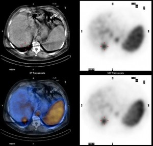
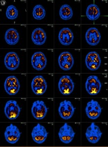
PET (Positron Emission Tomography) is rapidly becoming a major diagnostic imaging modality used predominantly in determining the presence and severity of cancers, neurological conditions, and cardiovascular disease. It is currently the most effective way to check for cancer recurrences. Studies demonstrate that PET offers significant advantages over other forms of imaging such as CT or MRI scans in diagnosing disease.
PET is considered particularly effective in identifying whether cancer is present or not, if it has spread, if it is responding to treatment, and if a person is cancer free after treatment. Cancers for which PET is considered particularly effective include lung, head and neck, colorectal, esophageal, lymphoma, melanoma, breast, thyroid, cervical, pancreatic, and brain, as well as other less frequently occurring cancers.