
Cerebrospinal Fluid Leak
A 41 year old Caucasian male with a history of hypertension presented to the emergency room with a 3 week history of worsening headache and neck stiffness. His neurologic exam was unremarkable for focal deficits. CT scan and MRI showed bilateral subdural collections suggesting chronic subdural hematomas due to intracranial hypotension. Radionuclide cisternography was performed with intra-thecal injection of In-111 DTPA through lumbar puncture, followed by planar imaging at 2 and 24 hrs and SPECT/CT imaging at 24 hrs.
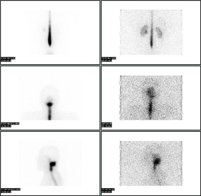
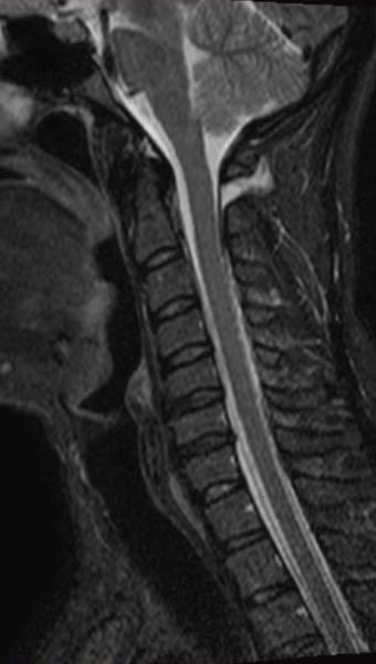
An abnormal focus of intense uptake was seen at the base of the skull on 2 hr and 24 hr planar images, without any discernable basal cistern or lateral ventricle activity or activity around the convexities. SPECT/CT localized the CSF leak to outside the thecal sac in the soft tissues between C1 and C2 towards the left of the midline. Subsequent MRI of the cervical spine confirmed the location and extent of CSF leak. After the placement of epidural blood path, patient reported dramatic improvement of his headaches.
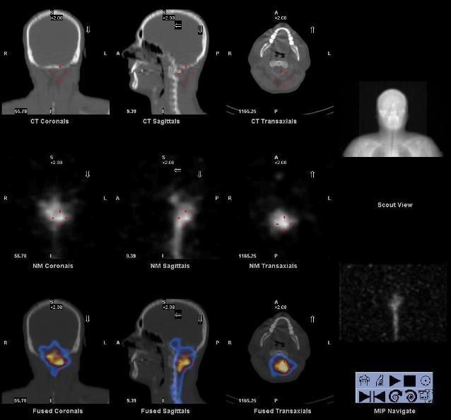
Radionuclide cisternography is a very sensitive for the detection of CSF leak. Once CSF leak is identified on planar images, SPECT/CT can be performed to localize the site and visualize other pathology.
This case was compiled by Dr. David He, BCM