
Metastatic Carotid Body Tumor
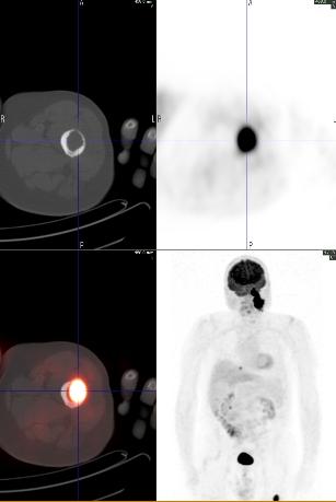
The patient is a 78 year old man with a history of a left neck glomus tumor diagnosed about 10 yrs ago, presented with complaints of worsening left thigh pain. X-ray showed a 2.4 cm lytic lesion in proximal left femur. CT-guided biopsy demonstrated metastatic neuroendocrine tumor.
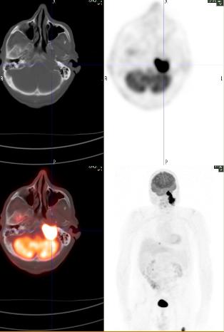
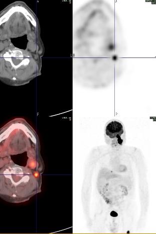
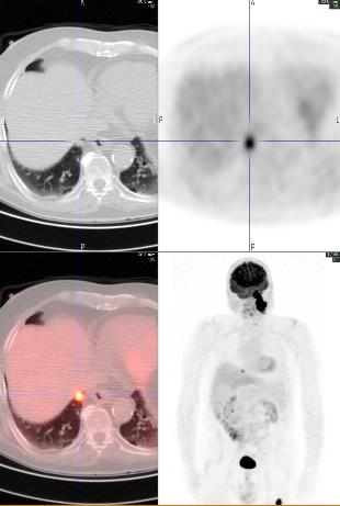
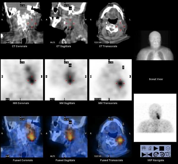
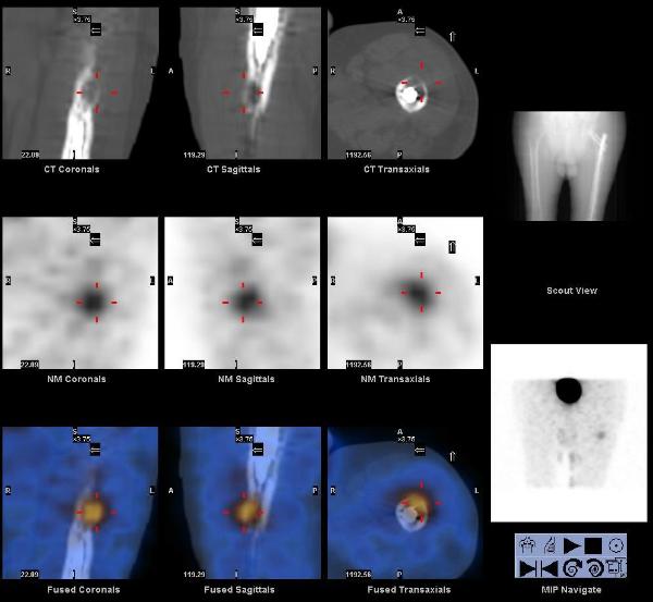
A whole body FDG PET-CT showed intense focal hypermetabolism (SUV max 26) in the large left neck mass extending from the base of the skull to the superior aspect of the hyoid bone, invading the left petrous temporal bone, and encasing the internal carotid artery and jugular vein. Hypermetabolism was also seen in left level IIb lymph nodes (SUV max 9.5), a 1.4 cm nodule in the right lung base (SUV max 6.4), and a lytic lesion in the left proximal femur (SUV max 18.0).
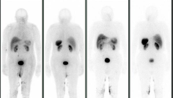
A subsequent In-111 Octreotide scan showed increased tracer uptake in the left neck mass and femur, but not in the cervical lymph nodes or lung nodule.
More metastatic lesions were seen on the FDG PET-CT compared to the Octreotide SPECT-CT. In the interim patient also had open reduction and internal fixation at the left femur.
Carotid body tumors are paragangliomas (extra-adrenal pheochromocytomas) arising from the carotid body. They arise close to or envelop the bifurcation of common carotid artery, usually in the sixth decade of life, and may be familial with autosomal dominant transmission in MEN 2 syndrome. They frequently recur after resection, many metastasize, and 50% ultimately prove fatal by direct invasion.
1. FDG PET imaging of paragangliomas of the neck: comparison with MIBG SPET. Macfarlane DJ, Shulkin BL, Murphy K, Wolf GT. Eur J Nucl Med. 1995 Nov;22(11):1347-50.
2. PET scan assessment of chemotherapy response in metastatic paraganglioma. Argiris A, Mellott A, Spies S. Am J Clin Oncol. 2003 Dec;26(6):563-6.
This case was compiled by Dr. Matthew R. Galfione, BCM