
Splenosis on SPECT-CT
A 60 yrs old male, smoker, with a past medical history of hypertension, and remote h/o post trauma splenectomy presented with symptoms of UTI. He was found to have microscopic hematuria which did not resolve with antibiotics and a CT scan of the abdomen was performed. It showed multiple enhancing soft tissue density masses in the abdomen and pelvis.
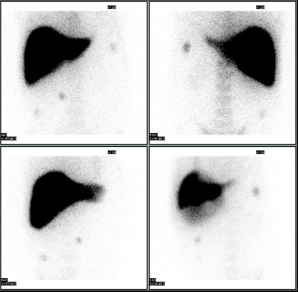
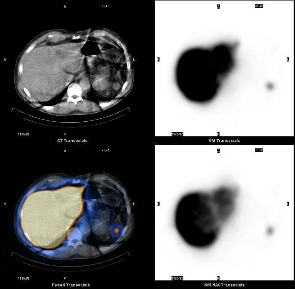
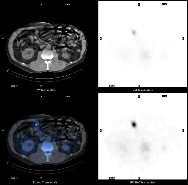
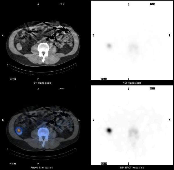
Splenosis was suspected and a liver-spleen scan was acquired after the injection of 8.7 mCi of Tc-99m sulfur colloid. Planar images showed multiple scattered foci of increased uptake in the abdomen and pelvis, consistent with splenosis. SPECT-CT images showed that all the enhancing lesions seen on the diagnostic CT corresponded to foci of increased uptake, consistent with splenic tissue.
1. Splenosis: 99mTc-labelled colloids provide the diagnosis in splenectomised patients. Franceschetto A, Casolo A, Cucca M, Bagni B. Eur J Nucl Med Mol Imaging. 2006 Sep;33(9):1102.
2. Splenosis presenting as multiple intra-abdominal masses mimicking malignancy.Kok J, Lin M, Lin P, Ngu C, Sam S, Loh C, Kociuba K. ANZ J Surg. 2008 May;78(5):406-7.
This case was compiled by Dr. Joji Varghese, MEDVAMC