
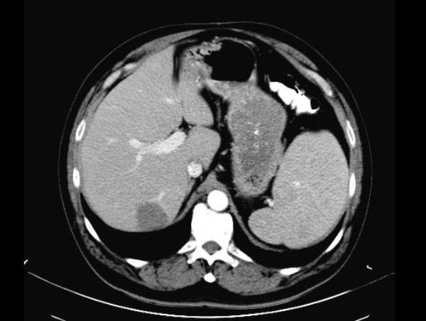
Hepatic Hemangioma
60 yrs old male presented with a 3.2 x 2.8 cm indeterminate liver mass seen in the right lobe, on the CT scan. Subsequent MRI study found characteristics atypical for a hemangioma.
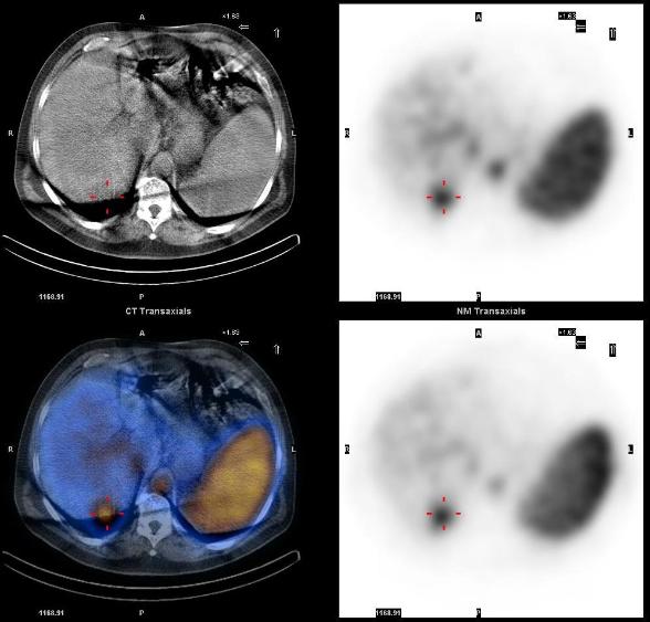
Tc-99m labeled RBC scan showed a focus of increasing uptake on the dynamic images (posterior projection), and focal intense uptake corresponding to the liver mass, on the SPECT-CT, characteristic of hemangioma.
1. Review of hepatic imaging and a problem-oriented approach to liver masses. Bennett WF, Bova JG., Hepatology. 1990 Oct;12, 761-75
This case was compiled by Dr. David He, BCM