
Metastatic Rectal Carcinoid
A 65 years old asymptomatic male who tested positive for fecal occult blood testing was found to have a rectal mass on colonoscopy, initially reported as an adenocarcinoma. Staging workup included a CT scan of the abdomen which showed a neoplasm in the liver, with neuroendocrine features on fine needle aspiration.
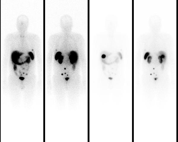
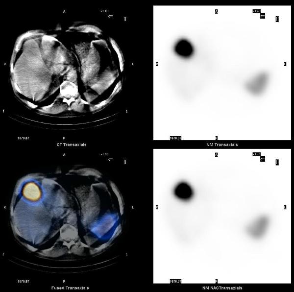
Planar 24 hr whole body In-111 octreotide scan images showed a small focus of mild uptake in lateral left lower chest, a large focus of intense uptake in the liver, and four distinct foci of mild to moderately increased uptake in the lower abdomen and pelvis.
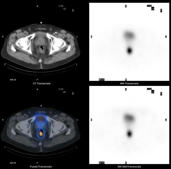
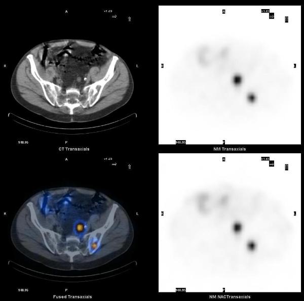
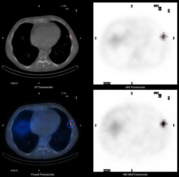
SPECT-CT images localized these abnormalities to: focal uptake in the rectal wall thickening, two presacral soft tissue nodules, the liver mass, and multiple bone metastasis (left ilum, L5 pedicle, and the left 6th rib).
The overall incidence of carcinoid tumor is increasing, partly due to improvements in diagnostic imaging. The best modalities for diagnosis and staging of carcinoids are In-111 Octreotide, I-123 MIBG and F-18 FDG PET-CT. SPECT-CT imaging helps in accurate localization of abnormal uptake seen on planar scintigraphy. Rectum is the third most common site of gastrointestinal carcinoid tumors. Rectal carcinoids are usually asymptomatic, carcinoid syndrome does not occur in these patients, and the incidence of metastases is low.
1. Anatomic and functional imaging of metastatic carcinoid tumors. Scarsbrook AF, Ganeshan A, Statham J, Thakker RV, Weaver A, Talbot D, Boardman P, Bradley KM, Gleeson FV, Phillips RR. Radiographics. 2007 Mar-Apr;27(2):455-77.
2. A proposed staging system for rectal carcinoid tumors based on an analysis of 4701 patients. Landry CS, Brock G, Scoggins CR, McMasters KM, Martin RC 2nd. Surgery. 2008 Sep;144(3):460-6.
This case was compiled by Dr. Sabeen Rahman, BCM