
Cuboid Fracture
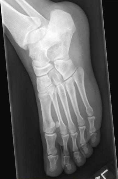
A 31 year old male presents with left ankle pain and swelling after a twisting injury during a basketball game. The pain radiates down to the anterior lateral proximal foot and is exacerbated when he plays basketball. X-rays of the left tibia/fibula, ankle and foot were unremarkable with no evidence of fracture or dislocation.
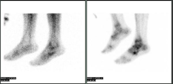
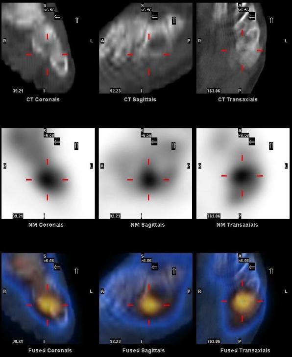
A three phase bone scan was performed which showed focal hyperemia on blood flow and blood pool images, and focal osteoblastic activity on the delayed images in the left mid foot. SPECT/CT acquired after the delayed planar images showed that the focal Tc-99m MDP uptake corresponds to a subtle hairline fracture in the cuboid bone (best seen on the saggital projection) on CT images.
Gnanasegaran G et al. Multislice SPECT/CT in Benign and Malignant Bone Disease: When the Ordinary Turns Into the Extraordinary. Seminars in Nuclear Medicine. Volume 39, Issue 6, November 2009, Pages 431-442.
This case was compiled by Dr. Niraj Patel, BCM