
Dislocated Hip Prosthesis
An 86 years old female with bilateral hip prosthesis presented with pain in the left lower extremity, radiating to the upper and medial thigh. The pain is worse with activity and is relieved with rest. No history of injury. Past medical history is significant for high blood pressure, high cholesterol, cerebrovascular accident, and osteoporosis. The left hip prosthesis has been revised several times in the past. Patient complains of pain with resisted flexion of left hip. There is no evidence of swelling or skin lesions. X ray of the left hip shows possible loosening and subluxation. A three phase bone scan was performed.
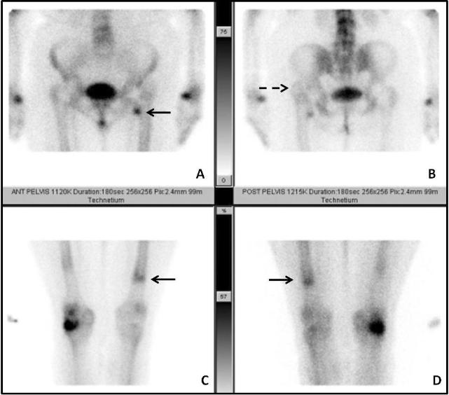
Blood flow and blood pool images (not shown) were significant for mild focal hyperemia in the left lesser trocanter. The delayed images show several signs of prosthesis dislocation. The head of the prosthesis is located in the acetabulum, but the femur has dislocated posteriorly and superiorly relative to the prosthesis.
Image A shows focal uptake in the left lesser trocanter, suspicious for fracture. Image B shows superior location of the left greater trocanter relative to the right hip. Images C & D show focal uptake at the distal end of the prosthesis from the downward dislocation of the prosthesis, relative to the femur.
Three phase bone scan is the standard of care for patients presenting with symptoms and sign suggestive of prosthesis complication. It is usually followed up with an In-111 WBC scan to evaluate for infection and sometimes with a third sulfur colloid bone marrow scan to confirm or rule out active infection.
Nuclear medicine and the infected joint replacement. Love C, Marwin SE, Palestro CJ. Semin Nucl Med. 2009 Jan;39(1):66-78.
This case was compiled by Dr. Amin Samarghandi, Excel Diagnostics