
Recurrent Paraganglioma
A 65 yrs old male was found to have extremely high blood pressure during work up for pacemaker placement. Subsequent biochemical studies revealed elevated urine and plasma cathecolamine metabolites. Patient has past medical history of right sided retroperitoneal paraganglioma resected 23 years ago.
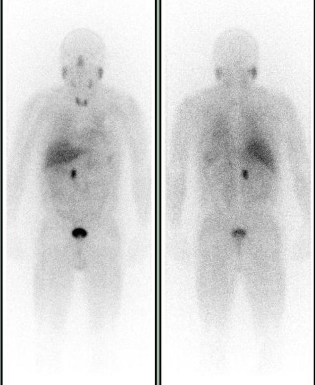
Suspected to have a recurrent paraganglioma or new pheochromocytoma, patient was referred for an MIBG scan to localize the tumor and possible metastasis. Whole body images show focal abnormal uptake in the right paramedian midabdomen.
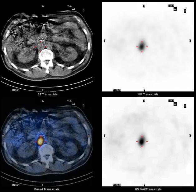
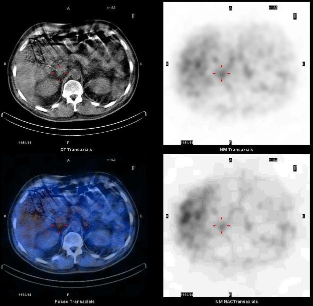
I-123 MIBG SPECT-CT demonstrates a focus of markedly increased uptake in the abdomen, corresponding to a soft tissue mass in the right retroperitoneal paracaval region just superior to the right renal vessels. Mild physiologic uptake is seen in the normal adrenals. The tumor and the right adrenal gland were surgically removed without complications. Pathology identified malignant paraganglioma in the tumor and the normal adrenal. MIBG SPECT-CT helped localize recurrent paraganglioma for surgical resection.
1. Detection and treatment of pheochromocytomas and paragangliomas: current standing of MIBG scintigraphy and future role of PET imaging. Havekes B, Lai EW, Corssmit EP, Romijn JA, Timmers HJ, Pacak K. Q J Nucl Med Mol Imaging. 2008 Dec;52(4):419-29.
2. Scintigraphic imaging of body neuroendocrine tumors. Intenzo CM, Jabbour S, Lin HC, Miller JL, Kim SM, Capuzzi DM, Mitchell EP. Radiographics. 2007 Sep-Oct;27(5):1355-69.
This case was compiled by Dr. David He, BCM