
Abdominal Abscess
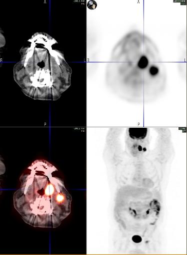
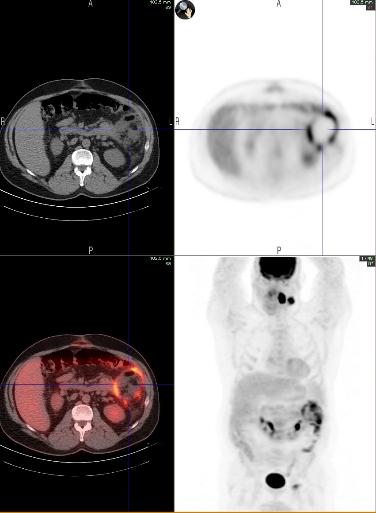
A 54 year old male with a history of hepatitis C, stage II colon cancer, status post left hemicolectomy presented for follow-up of a newly diagnosed left tonsillar squamous cell carcinoma. A whole body FDG PET/CT was preformed for staging, two months after the surgery for colon cancer. Focal hypermetabolism was seen in the left tonsillar mass and left level II lymphadenopathy. Incidentally noted increased uptake in the left upper quadrant of the abdomen was thought to be related to post surgical changes at the site of prior bowel anastomosis. Patient also has history of recurrent MRSA infections (subcutaneous abscess in the lower extremity, groin, and axilla).
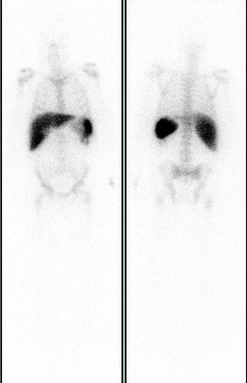
Soon after initiating chemotherapy patient was admitted for fever with chills and was found to have MRSA bacteremia. In-111 labeled leukocyte (WBC scan) study was performed with 657 microcuries to identify the source of infection. Planar images show ill defined focal uptake in the left upper quadrant of the abdomen, abutting the spleen.
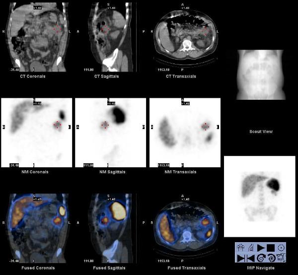
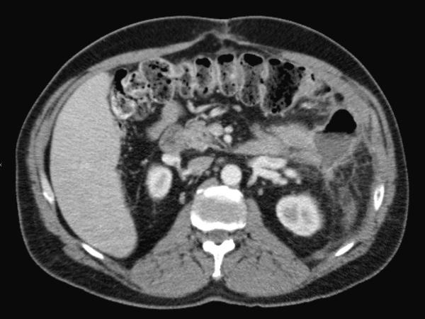
The SPECT/CT images localize the focus to the splenic flexure of colon, at the site of prior bowel anastomosis, post left hemicolectomy. This abnormal accumulation of In-111 labeled WBCs was suspicious for active infection. A follow up contrast enhanced CT scan of the abdomen showed an abscess in this region, that was drained.
Early investigation of hybrid SPECT/CT imaging technology in localizing infection has been very promising. This could lead to a greater acceptance of this technology in a wide variety of additional clinical roles as well.
Bybel, B et al. SPECT/CT imaging: clinical utility of an emerging technology. Radiographics. 2008 Jul-Aug;28(4):1097-1113.
Heiba, S et al. The diagnostic confidence of SPECT & SPECT/CT in Indium-111 leukocyte scintigraphy. J Nucl Med. 2007; 48 (Supplement 2):63P.
This case was prepared by Dr. Raj R Chinnappan BCM