
Diabetic Foot Infection
A 75 year old male with a history of type II diabetes mellitus presented for further evaluation of an ulcer on the distal lateral aspect of the right foot. It had worsened with conservative treatment in the last few months with symptoms of swelling, tenderness, and drainage. Underlying osteomyelitis was suspected.
Radiographs of the foot demonstrated midfoot arthrosis and old postsurgical changes from a 4th ray amputation at the level of the distal 4th metatarsal, but no evidence of osteomyelitis.

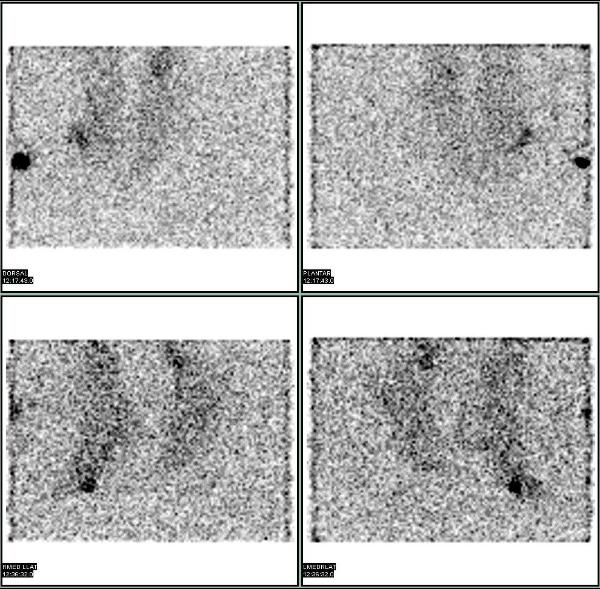
An In-111 labeled leukocyte study (WBC scan) was performed after the intravenous injection of autologous WBCs labeled with 540 microcuries of In-111. Planar images demonstrated a small focus of mild to moderately increased uptake in the region of the ulcer, suggesting active infection. The planar images lack the anatomical landmarks / resolution to tell whether the uptake is in the soft tissues or bone.
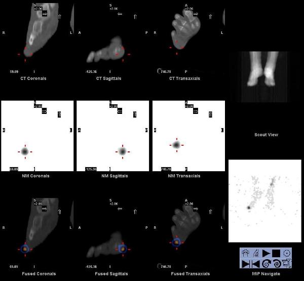
SPECT-CT images of the foot were acquired. The images localize the uptake to the soft tissues and osteomyelitis was ruled out. Fusion of functional SPECT data with structural CT data provides anatomic localization. While this rapidly emerging hybrid imaging technology is of clear benefit in the diagnosis of osteomyelitis in diabetic foot infections, the broader clinical implications from such improved anatomical accuracy are limitless.
Bar-Shalom R, et al. SPECT/CT using 67Ga and 111In-labeled leukocyte scintigraphy for diagnosis of infection. J Nucl Med. 2006 Apr;47(4):587-94.
Horger M, et al. Added value of SPECT/CT in patient suspected of having bone infection: preliminary results. Arch Orthop Trauma Surg. 2007 Apr; 127 (3):211-221.
This case was compiled by Dr. Raj R Chinnappan, BCM